- Share
- Share on Facebook
- Share on LinkedIn
OLIMPICS events
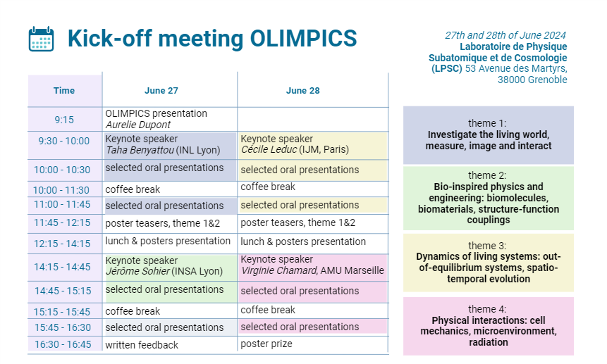
From 27 June 2024 to 28 June 2024
Grenoble - Presqu'île
Full program here
Start of the event (June 27 and 28): 9:15
End of the event (June 27 and 28): 16:45
Posters presentation (June 27 and 28): from 12:15 to 14:15
Poster Prize : June 28 at 16:30
Keynote speakers :
June 27 : Taha Benyattou (INL Lyon, Theme 1), Jérôme Sohier (INSA Lyon, Theme 2)
June 28 : Cécile Leduc (IJM, Paris, Theme 3), Virginie Chamard (AMU Marseille, Theme 4)
Abstracts :
Taha Benyattou (INL, UMR5270, 69622 Villeurbanne, France)
Title : Piégeage optique à l’aide de cavité de cavité à mode de Bloch lent : Depuis la manipulation de nano-objets vers la mesure le mesure de déformabilité de cellules uniques
Abstract : L’utilisation de nanostructure photonique a ouvert depuis plus d’une décennie de nouveaux horizons pour le piégeage optique. Nous montrerons dans un premier temps comment l’utilisation du champ proche a permis le piégeage et la manipulation de nanoparticules. La seconde partie de cet exposé sera dédiée aux applications dans le domaine du vivant. En particulier, nous détaillerons l’approche que nous avons développée pour la mesure tout optique de la déformabilité de cellules uniques.
Projets ANR Baltrap (ANR 2010 NANO 002 01) et CellDance (ANR-21-CE09-0011).
Jérôme Sohier (MATEIS INSA Lyon, France)
Title : Biomimetic hydrogels: structural versatility as a tool for modulating cellular interaction and reproducing complex tissues.
Abstract : Hydrogels are valuable tools for mimicking the extracellular matrix structure and qualities due to their biocompatibility, mechanical properties, permeability to oxygen and nutrients, and high water content. They can be designed, tuned, and formulated to either favor and reproduce interactions with cells or to mimic the structure and function of complex tissues. To explore these potentials, we have developed synthetic hydrogels with versatile and standardized properties that can replicate a wide range of tissue viscoelastic properties.
In an initial investigation, we evaluated their potential for cellular interaction and determined their ability to modulate human skin and muscle cell behavior based on the hydrogels' inherent structural and mechanical properties. Through innovative formulations, we then further elucidated structure-function relationships in 3D culture models of muscle tissue.
We then explored the possibility of reproducing the structural complexity and unique mechanical behavior of human vocal folds. From precise characterization of native human tissues, we mimicked both the collagen network and the elastin/ground substance of the extracellular matrix with polymer fibers and hydrogel composition. This approach allowed us to successfully replicate the non-linear, anisotropic, and viscoelastic mechanical behavior of the vocal folds.
These examples will be presented, with an emphasis on the importance of structure-function relationships in biomimetic designs.
Cécile Leduc (Institut Jacques Monod, Paris, France)
Title : Mechanics and dynamics of single intermediate filaments
Abstract : Intermediate filaments (IF) are involved in key cellular functions including polarization, migration, and protection against large deformations. These functions are related to their remarkable ability to extend without breaking, a capacity that should be determined by the molecular organization of subunits within filaments. However, this structure-mechanics relationship remains poorly understood at the molecular level. I will present how, using super-resolution microscopy (SRM), we show that vimentin filaments exhibit a ~49 nm axial repeat both in cells and in vitro. Using an SRM compatible stretching device, we also provide evidence that the extensibility of vimentin is due to the unfolding of its subunits and not to their sliding, thus establishing a direct link between the structural organization and its mechanical properties. In a second part, I will show recent results on the in vitro reconstitution of vimentin assembly dynamics. We show that vimentin filaments can spontaneously break without cofactors or post-translational modifications, but this fragmentation limits assembly only at very long-time scale (>18h). We also uncovered the mechanism responsible for fragmentation which involves subunit exchange. Our results show how vimentin self-repair to protect their integrity and provide new insights into the physical understanding of the intermediate filament length regulation.
Virginie Chamard (Institut Fresnel Marseille)
Title : Physico-chemistry insights in calcareous biomineralisation gained from advanced x-ray and optical microscopy approaches
Abstract : Biomineralization integrates complex physical and chemical processes bio-controlled by living organisms through ionic concentration regulation and organic molecules production. The capability to tune, from ambient conditions crystallisation, the structural, optical and mechanical properties of hard tissues motivates extensive research to transfer biomimetic approaches into material science studies. This urges a detailed understanding of the underlying processes at play in Nature.
Our work focuses specifically on the mechanisms underlying the production of calcareous hard tissues in marine species (oysters, corals, coccoliths, etc…). While it is clear that non-classical crystallisation processes govern the formation of most of these biominerals, major questions are pending regarding the involved physico-chemical pathways. These include the identification of - possibly - several metastable precursors and the nature of their successive transitions.
Our strategy is based on the investigation of early-mineralized units, located at the growth front of the biomineralizing tissue, which we characterize with highly sensitive and highly spatially resolved optical or x-ray microscopy approaches. In particular:
- To monitor the different chemical states (e.g., amorphous or crystallized carbonates, organics molecules), a highly sensitive coherent Raman microscopy approach is developed. It allows mapping molecular bond concentrations and demonstrates suitability for the in vivo imaging of a growing biomineral (e.g., Pinctada margaritifera oyster shell).
- To address the crystallization process, we make use of x-ray scanning nano-diffraction at novel 4th generation synchrotron source. It provides nanoscale spatially resolved and crystalline-sensitive study of a biomineralizing animal (Crassostrea gigas shell and Stylophora pistillata coral).
In this presentation, I will show recent results, which bring new insights in calcareous biomineralization pathways.
Date
Localisation
Grenoble - Presqu'île
Laboratoire de Physique Subatomique et de Cosmologie (LPSC, 53 Av. des Martyrs, Grenoble)
- Share
- Share on Facebook
- Share on LinkedIn